
Microscopic root tip resection
Surgical root tip resections (WSR) are carried out at the My Dent and Face practice when a tooth with an inflamed root tip needs to be saved.
When is a root tip resection necessary?
The following indications are conceivable for a root tip resection:
- Conventional root canal treatment and subsequent revision were unable to resolve the inflammatory processes at the root tip.
- X-ray diagnostics show changes in the area around the root tip that require a detailed histological examination.
- The position or shape of the tooth roots is unusual, so the root filling cannot be completely inserted.
- There are tooth and/or root injuries/root fractures.
- Cysts were identified around the root tip.
- There is suspicion of a tumor.
- The root canal can no longer be prepared due to complications from previous treatments.


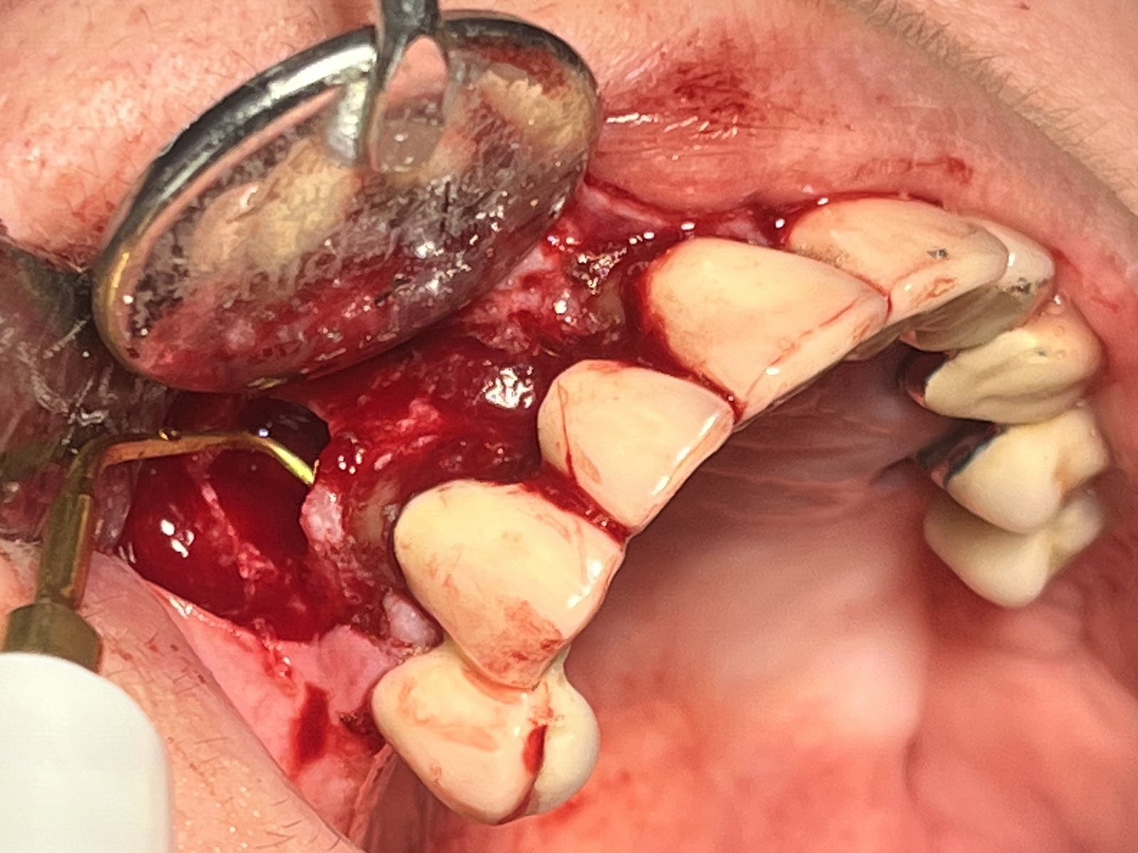
Radicular cysts on teeth 12 and 13; tooth 12 was extracted through the bridge without removing the bridge structure with the help of a marginal incision. Tooth 13 has a radicular cyst; a root resection was performed to preserve the tooth. With the help of piezosurgery, the root canal of 13 was expanded retrogradely so that there was no recurrence. The two bone defects were filled with cast substitute material.
Root tip resection – FAQ
The surgical procedure
The operation is usually completely painless under local anesthesia.
Every root tip resection in My Dent and Face is preceded by a thorough diagnosis. Often – especially in more difficult cases – it makes sense to look at the root tip in great detail on a three-dimensional X-ray image (DVT – digital volume tomography). If other areas of inflammation in the maxillary sinus arise from chronically inflamed teeth, this can best be seen on a 3D image. This is very important for comprehensive and sustainable treatment success.
The treatment process for root tip resection
- Severing of the gums and periosteum
- Exposure of the bone in the area of the tooth root
- Removal of the bone tissue with special milling machines or with the help of ultrasound-assisted surgery (piezosurgery – a modern, particularly gentle procedure that is used in the My Dent and Face practice clinic)
- Shortening of the branched extensions of the root canal, the root tip, by usually around 2-3 mm
- Expansion of the root canal
- Disinfection of the root canal using rinsing solutions or – even more thoroughly – using the dental laser.
- Drying of the root canal
- Fill the prepared root canal with gutta-percha (a rubber-like material). If access to the root tip via the tooth crown is not possible, a filling can also be done from the other side, i.e., from the root tip.
- Suturing the soft tissue over the wound
- The procedure is rechecked with an x-ray.
- Temporary filling of the treated tooth
- Final filling of the treated tooth after the wound has healed.
In some cases – especially with the posterior teeth of the upper jaw – it may make sense to carry out the operation from the palate. The teeth there are multi-rooted, with the individual roots often being far apart from each other.
After the operation, you will not be allowed to eat or drink until the anesthesia has worn off. Coffee and black tea, as well as major physical exertion, are only permitted 24 hours after the procedure. Use cooling pads to prevent swelling and bruising at home. Impeccable oral hygiene – being very careful in the area of the operated tooth – is also essential for successful healing.
Another X-ray check will be carried out after three or at least six months.